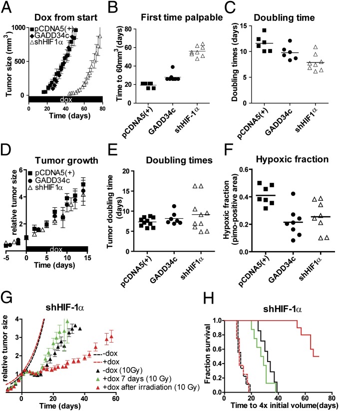
Fig. 4.
PERK/UPR and HIF deficiency delays tumor formation. (A) Tumor growth of s.c. implanted isogenic HCT116 cells targeting HIF-1α (i.e., shHIF-1α), GADD34c, and control cells [pCDNA5(+)] preexposed (24 h) to doxycycline (dox) were injected into doxycycline receiving nude mice [mean ± SEM, pCDNA5(+), n = 6; GADD34c, n = 8; shHIF-1α, n = 8]. (B) Palpability (>60 mm3) of tumors. (C) Doubling time of individual tumors. (D) After establishment (150 mm3), mice received doxycycline in their drinking water [mean ± SEM; pCDNA5(+), n = 10; GADD34c, n = 7; shHIF-1α, n = 10]. (E) Doubling time of the individual tumors. (F) Tumors were harvested after 7 d doxycycline, and hypoxia was assessed by pimonidazole immunohistochemistry of the viable tumor tissue. (G) Mice with established tumors (150 mm3) received doxycycline in drinking water. Growth (dotted lines) and regrowth after irradiation (t = 0) was followed over time in animals that received no doxycycline (−dox, n = 7) or doxycycline from day −4 to day +3 [+dox 7 d (10 Gy), n = 8] or doxycycline after irradiation [+dox after irradiation (10 Gy), n = 10]. (H) Kaplan–Meier survival for tumors in Fig. 4 D and G to with the endpoint of reaching four times the initial volume.

