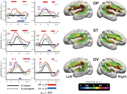
Fig. 3.

Discriminative responses to auditory stimuli in premature infants. (Right) Surface-based topographic color map of the HbO response at the peak of the hemodynamic response for the three conditions. (Left) HbO time courses for the youngest and oldest subsets of six infants, recorded over left Broca’s area (ch 12), left planum temporale (ch 18), left superior temporal gyrus (ch 20), and their counterlateral right channels (ch 5, ch 8, and ch 6, respectively). The colored rectangles indicate the time windows during which the deviant conditions differ significantly from the standard condition using cluster-based statistics over the whole group. The direction of the effect is given by the location of the bar under or above the x-line. Preterm infants discriminate the change of phoneme, notably in the left inferior frontal region. This effect was stable across our age range (28–32 wGA).
