


Fig. 1.
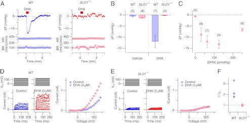
DHA lowers blood pressure in wild-type mice and enhances SMC BK channel currents. (A) Representative recordings of changes in arterial blood pressure (ΔP) , heart rate (HR), and breathing rate (BR) in wild-type (WT; Left) and Slo1 knockout (SLO1−/−; Right) mice by injection of DHA (62.5 μmol/kg) at the times indicated by the red bars. (B) Analysis of peak deflections of blood pressure in wild-type and SLO1−/− mice on injection of DHA and the vehicle (0.1% Tween-20 in 154 mM NaCl). Blood pressure values in WT mice before and after DHA injection were significantly different (P < 0.05) but no change was observed with the vehicle. In SLO1−/−, neither DHA nor the vehicle produced any change in blood pressure. (C) Dose dependence of the peak blood-pressure lowering effect of DHA. (D) Representative whole-cell currents (Left) from an isolated aortic SMC from a wild-type mouse elicited by voltage pulses before (blue) and after (red) application of DHA (3 µM). Peak current-voltage (IV) curves are also shown (Right). (E) Representative whole-cell currents (Left) and IV curves (Right) from an isolated aortic vascular SMC from a SLO1−/− mouse. (F) Fractional increase in the peak outward current size at 80 mV by DHA (3 µM for 4 min; see Fig. 2D) in WT and SLO1−/− mice. Fractional increases in the two groups are statistically different (P < 0.008). n = 5.