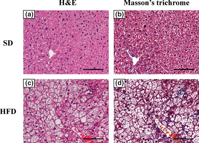
Figure 2.

Representative liver histopathology in C57BL/6J mice fed standard diet (SD) or high-fat diet (HFD) for 24 weeks. Haematoxylin and eosin (H&E) staining and Masson's trichrome staining of liver in SD-fed mice (a, H&E staining; b, Masson's trichrome staining, 200× magnification). The HFD-fed mice showed steatosis without fibrosis (c, H&E staining; d, Masson's trichrome staining, 200× magnification). Scale bars = 100 μm.
