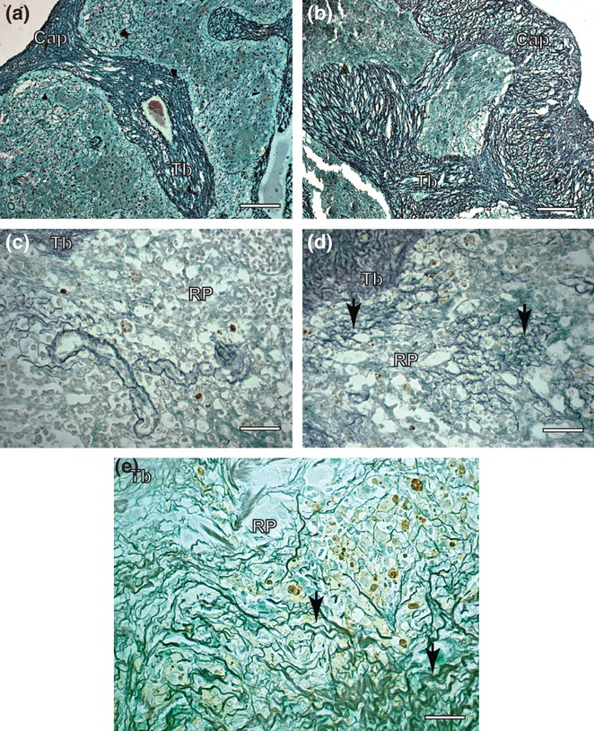
Figure 2.

Spleen sections of control and symptomatic dog naturally infected with Leishmania (L.) chagasi. (a) Control dog: observe capsule and trabecules system composed by reticular fibers Ammoniacal silverstaining (Bars = 32 μm); (b) Infected dog: note intense argyrophilic fibers deposition in the capsule and trabecules. Ammoniacal silver-staining (Bars = 32 μm); (c) Control dog: observe inside red pulp reticulin fibers as a delicate network. Ammoniacal silver-staining (Bars = 16 μm); (d,e) Infected dog: inside of red pulp besides in higher intensity, the fibers were thicker, dense and coiled than controls, forming a conspicuous network (black arrows). Ammoniacal silver-staining (Bars = 16 μm); Cap, capsule; Tb, trabecule; RP, red pulp.
