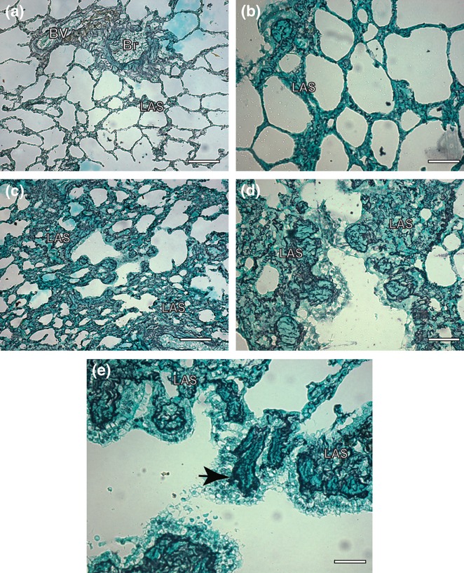
Figure 4.

Lung sections of control and symptomatic dog naturally infected with Leishmania (L.) chagasi. (a,b). Control dog: observe a delicate reticular fibers network within the alveolar septa Ammoniacal silverstaining (Bars = 32 μm and Bars = 16 μm, respectively); (c,d) Infected dog: note intense argyrophilic fibers deposition in the within the alveolar septa associated to an intense chronic inflammatory response. Ammoniacal silver-staining (Bars = 32 μm and Bars = 16 μm, respectively); (e) Infected dog: besides in higher intensity, the fibers were thicker, dense and coiled than controls, forming a conspicuous network (black arrow). Ammoniacal silver-staining (Bars = 16 μm); LAS, lung alveolar septa; Br, bronchioles; BV, blood vessels.
