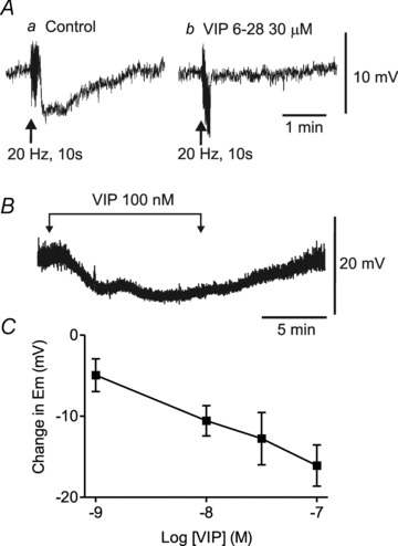
Figure 11. Non-nitrergic, non-purinergic (NNNP) hyperpolarization is abolished by VIP6–28 and mimicked by exogenous VIP.

A, sample traces showing the post-stimulus hyperpolarization accompanying 20 Hz EFS for 10 s (Aa). This hyperpolarization is blocked following 17 min superfusion with VIP6–28 (30 μm; Ab). B, sample trace showing the hyperpolarization elicited with superfusion of 100 nm VIP. C, summary graph of the concentration-dependent effects of VIP (1–100 nm, n = 3–7) on membrane potential (Em). Shown are mean values ± SEM. Atropine, guanethidine, 1 μm MRS2500 and 100 μm l-NNA were present throughout.
