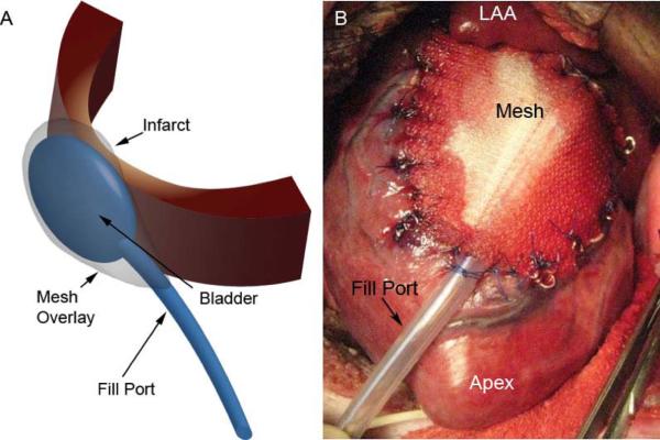
Figure 1. Restraint Device.

Panel A depicts a schematic drawing of the restraint device used in this study. A bladder type catheter is placed over the infarct between the epicardium and a mesh sutured to the heart surface. External filling is performed via an exteriorized fill port. Panel B is an intraoperative photograph of the implanted device sutured to the LV epicardium directly over the infarct region. MRI compatible markers are sutured to the edge of the mesh and epicardium to delineate the infarct on MR imaging.
