
Figure 2. The Effect of Device Filling on LV Geometry.
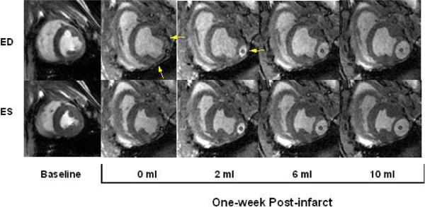
Mid-ventricular end-diastolic (ED) and end-systolic (ES) images at baseline and one week after infarct. From baseline to one-week post infarct the end diastolic volume (EDV) and end systolic volume (ESV) have increased significantly. Progressively increasing device volume from 0 ml to 10 ml decreases the infarct bulging and alters the infarct and LV geometry. Arrows in 0ml image denote the infarct region and in 2ml image point to the device.