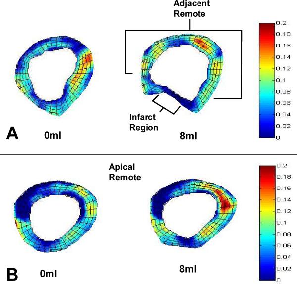
Figure 4. Color Map of Regional Maximum Principal Strain Pre and Post Device Filling.
(A) Device filling (8ml) altered both the geometry and strain in the basal region where the device was positioned. The septum and posterior wall had increased strain while the infarct region strain diminished. (B) The apical remote geometry was less affected by device filling while strain was significantly altered.

