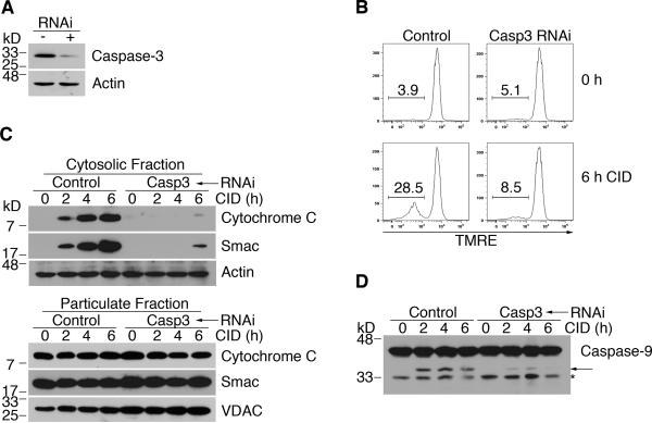
Figure 5. Knockdown of caspase-3 inhibits mitochondrial disruption induced by activation of caspase-9.
(A) Control H9-iCasp9 cells and cells expressing siRNA to silence caspase-3 were used for Western blot for casapse-3 or actin. (B) Cells as in (A) were treated with 10 nM CID and TMRE staining was performed at the indicated times to measure mitochondrial membrane potential. (C) Cells were fractionated at the indicated times. Western blotting for cytochrome c and Smac was performed. Membranes were re-probed with anti-actin or anti-VDAC for equal loading controls. (D) Cells were treated with CID and lysed for Western blotting at the times indicated. Arrow denotes processed caspase-9. Asterisk indicates degraded or non-specific protein. Results are representative of at least two experiments.

