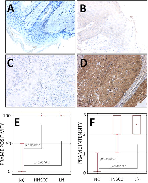
Figure 2. PRAME expression in normal control (NC) oral mucosa obtained from normal donors (NC), HNSCC lesions or metastatic lymph nodes (LN).

(A) Isotype Ab control in a specimen of NC mucosa (× 200); (B) No or only weak expression of PRAME in a specimen of NC mucosa (× 200); (C) Negative (isotype Ab) control in a specimen of HNSCC (× 200); (D) Strong expression of PRAME in a representative specimen of HNSCC (× 200); (E) PRAME expression (% POSITIVE CELLS) in NC vs. HNSCC or LN, respectively. All HNSCC and LN were scored at >75% positivity; (F) PRAME expression (INTENSITY, measured as described in Methods) in NC vs. HNSCC or LN, respectively flow cytometry.
