Figure 7.
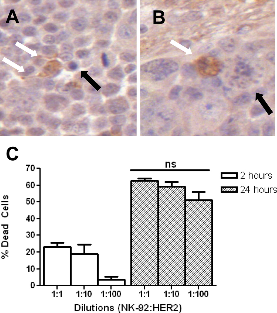
HER2-specific NK-92 cells accumulate at the tumor and have preserved function. A, IHC for granzyme B highlights an NK-92 cell releasing granzyme B into the surrounding extra-cellular space (white arrows). An adjacent apoptotic tumor cell can be seen (black arrow). B, granzyme B-containing NK-92 cell (white arrow) causing apoptosis in a tumor cell (black arrow). At 24 hrs, a 1:100 ratio of effector:tumor cells is statistically no different (p > 0.05) in causing tumor cell lysis than higher starting ratios, C. This is the ratio of effector-to-tumor cells that was achieved in vivo in group 3.

