Figure 1.
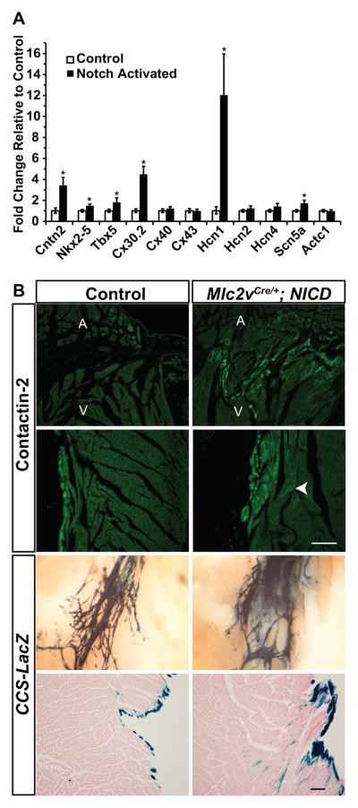
Activation of Notch Signaling Upregulates Conduction System-Enriched Genes. (A) RT-qPCR of conduction-enriched genes in ventricular samples from Mlc2vCre/+; NICD mice with transcript levels normalized to control littermates. Whereas there was no change in expression of the pan-cardiac marker Actc1 (alpha-cardiac actin), Notch activation up-regulates conduction-enriched transcription factor, gap junction, and ion channel genes. (B) Immunohistochemistry for the conduction-specific protein Cntn2 (green) demonstrates ectopic expression in Notch activated hearts when compared with control, including within the right ventricle near the atrioventricular junction and in the interventricular septum near the right bundle. The arrow demonstrates Cntn2-expressing cells within the interventricular septum in a region distant from the endocardium. Whole mount analysis of CCS-LacZ expression similarly demarcates ectopic conduction tissue along the right bundle with a blurring of the boundary between conduction and chamber myocardium in Notch activated hearts. Eosin stained sections from Notch activated hearts along the left side of the interventricular septum demonstrate ectopic CCS-LacZ expression broadly in the subendocardial region. Scale bars = 100μM, whole mount CCS-LacZ images are at 6.3× magnification. A=atrium, V=ventricle. Control mice are NICD. N=3 each genotype. Group comparison was performed using a Student’s unpaired 2-tailed t-test. *p<0.05

