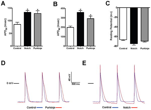
Figure 4.
Notch Converts the Cellular Electrophysiology of Newborn Ventricular Cardiomyocytes to a Purkinje-like Phenotype. Comparison of the action potential duration (APD) at 50% (A) or 90% (B) repolarization in newborn cardiomyocytes isolated from Cntn2-EGFP mice, which identifies endogenous Purkinje fibers. EGFP− chamber cardiomyocytes (white bars, n=9) have a significantly shorter APD50 and APD90 when compared with EGFP+ Purkinje cells (grey bars, n=8). Notch activation in EGFP− chamber cardiomyocytes (black bars, n=10) results in a significant prolongation of both the APD50 and APD90 towards a Purkinje-like phenotype. (C) The resting membrane potential was not significantly different between chamber cardiomyocytes, Purkinje cells, and Notch activated cells though there was slight membrane hyperpolarization in Purkinje cells and with Notch activation. (D) Superimposed action potentials from a control lacZ-infected (blue) and EGFP+ Purkinje cell (red). (E) Superimposed action potentials from a control lacZ-infected (blue) and Notch activated cell (red) demonstrate Notch-induced changes in the action potential morphology similar to those in Purkinje cells. Group comparison was performed using a one-way ANOVA and Newman-Keuls test for post-hoc analysis. *p<0.05

