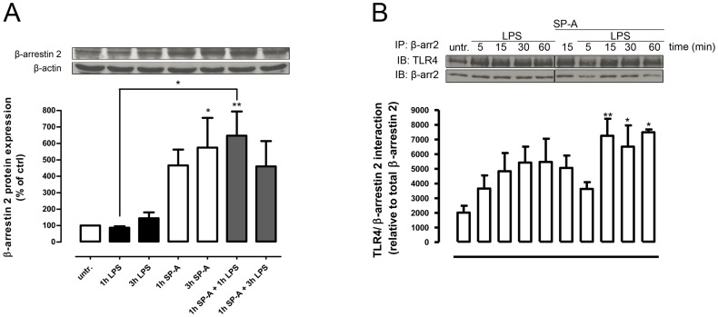
Figure 4. SP-A enhances β-arrestin 2 protein expression and LPS-induced β-arrestin 2/TLR4 interaction in primary AM.
A, Western blot of total β-arrestin 2 protein expression in primary rat AM treated with LPS (100 ng/ml, 1 h and 3 h), SP-A (40 µg/ml, 1 h and 3 h) or both (SP-A 1 h plus LPS 1 h or 3 h) as indicated. Equal amounts of whole cell lysates were subjected to SDS-PAGE and immunoblotted for β-arrestin 2 and β-actin. Data of at least four independent experiments were normalized to β-actin, basal β-arrestin 2 expression in untreated cells was set 100%, and calculated data were statistically analyzed by two-way ANOVA with Bonferroni's post test (mean ± SEM). *p<0.05; **p<0.01. B, Immunoprecipitation (IP) of β-arrestin 2 and TLR4 immunoblot (IB) from rat AM lysates treated with LPS (100 ng/ml), SP-A (40 µg/ml), or both as indicated. Data of seven independent experiments were analyzed by one-way Anova with Dunett's posttest (mean ± SEM). *p<0.05; **p<0.01 (versus untreated control).

