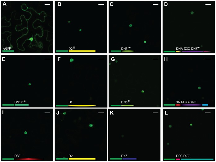
Figure 5. Localisation of GFP-tagged CRN C-termini.
A) shows localisation of free GFP. B-L) show a diverse range of GFP-tagged CRN C-termini. B = 1_719, C = 11_767, D = 12_997, E = 20_624, F = 32_283, G = 33_10, H = 36_259, I = 60_274, J = 79_188, K = 83_152, L = 105_26. All tested CRN fusions localise to the nucleus of the cell. Different subnuclear localisations can be observed for some CRNs (B, G, J, L). The domain organisations of the C-termini are represented as fused to GFP (green rectangle) for each image. NLS are predicted to be present in the genes marked with *. Scale bar = 25 µm.

