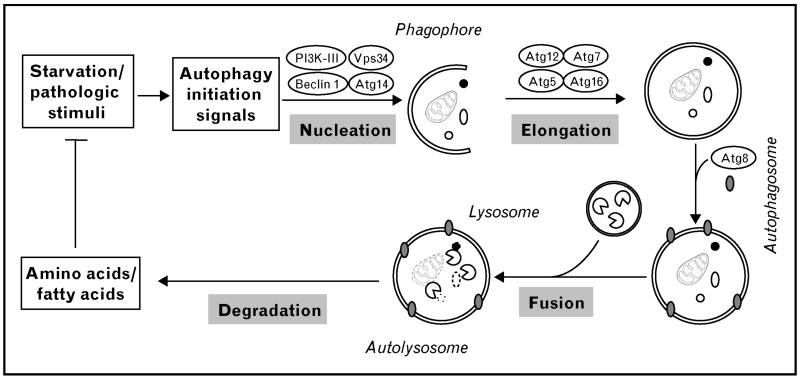
Figure 1. Schematic of autophagy pathways.
A variety of stimuli, including both physiological (e.g., starvation) and pathologic stresses, trigger cardiac autophagy. The first step in the process is nucleation. Classically, a complex composed of class III PI3K and Beclin 1 complex is involved. This complex initiates formation of an isolated double membrane and subsequent membrane elongation. The membrane then fuses upon itself, forming the distinctive double-membrane autophagosome. During the following fusion step, autophagosomes ultimately fuse with lysosomes, culminating in cargo degradation. The end-products of autolysosome cargo degradation provide both fuel and elemental building blocks to preserve vital cellular functions and remove toxic cellular elements. ATG, autophagy-related genes; PI3K-III, class III phosphatidylinositide-3-kinase; Vps34, vacuolar protein sorting 34.

