
FIGURE 2. The epidermal basal lamina, and neuronal development.
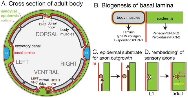
(A) Cross-section of adult body showing epidermis and adjacent tissues and structures. The external surface is covered by the cuticle; the internal face is covered by basal lamina (BL, red). Neurons (brown) reside on the epidermal side of the BL; the positions of the major nerve tracts relative to the dorsal and ventral epidermal ridges are shown. (B) Biogenesis of BL. The majority of known BL components are synthesized in muscles; the composition of the muscle-adjacent BL is likely distinct from that of non-muscle adjacent BL. (C) Developing axons appear to use the epidermis rather than the BL as substrate, as they are seen to pass on the epidermal side of other axons (R. Durbin, Ph.D thesis). (D). Sensory axons have a close relationship with the epidermis. Mechanosensory processes become progressively confined within the epidermis and secrete a specialized ECM, the mantle.