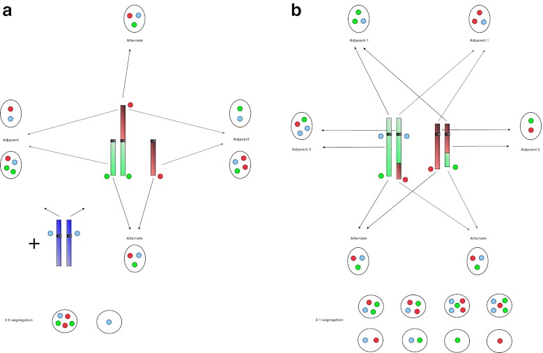
Fig. 1.
Sperm FISH patterns according to the type of translocation and the segregation modes. a Robertsonian translocations: one green and one red probes recognize the respective telomeric regions of the two acrocentric chromosomes involved. A third blue probe marks the centromere of another non-acrocentric chromosome, not involved in the translocation, in order to exclude diploid cells from the count. The alternate mode corresponds to the segregation of the two non translocated chromosomes in one sperm and of the translocated one in another sperm. The adjacent mode corresponds to the segregation of the translocated chromosomes with either one of the two normal ones. The 3:0 segregation mode leads to the segregation of the three involved chromosomes in one sperm, and none of them in another sperm. b Reciprocal translocations: one green and one red probes recognize the respective telomeric regions of the two chromosome arms involved, and a third blue probe marks the centromere of one of those two chromosomes. The alternate mode corresponds to the segregation of the two normal chromosomes in one sperm and of the two translocated ones in another sperm. The adjacent-1 mode corresponds to the segregation of one normal and one translocated chromosomes of each chromosomal pair in a sperm. The adjacent-2 mode corresponds to the segregation of the normal and the translocated chromosomes of the same chromosomal pair in a sperm. The 3:1 segregation mode leads to the segregation of three chromosomes in one sperm and the remaining chromosome in another sperm. In each case, the chromosomal content gives a specific combination of FISH signals

