
Figure 8.
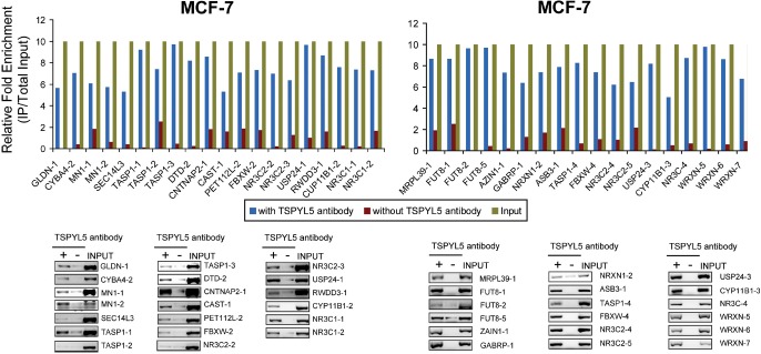
ChIP Assays That Demonstrate the Binding of TSPYL5 to Areas of Genes That Contained the Putative TSPYL5 DNA Binding Motif. ChIP was performed in MCF-7 cells with anti-TSPYL5 antibody. In each case the top panel shows the results of qRT-PCR demonstrating the relative enrichment of TSPYL5 DNA motif sequences after immunoprecipitation with anti-TSPYL5 antibody (blue) as compared with no antibody (IgG only) (orange). Sequences of primers utilized to perform these assays are listed in Supplemental Table 2. At the bottom of each figure are ChIP results after PCR amplification shown relative to input on a 1.0% agarose gel. Some genes contained more than one TSPYL5 DNA binding motif, so they were labeled as “-1”, “-2”, “-3” etc.