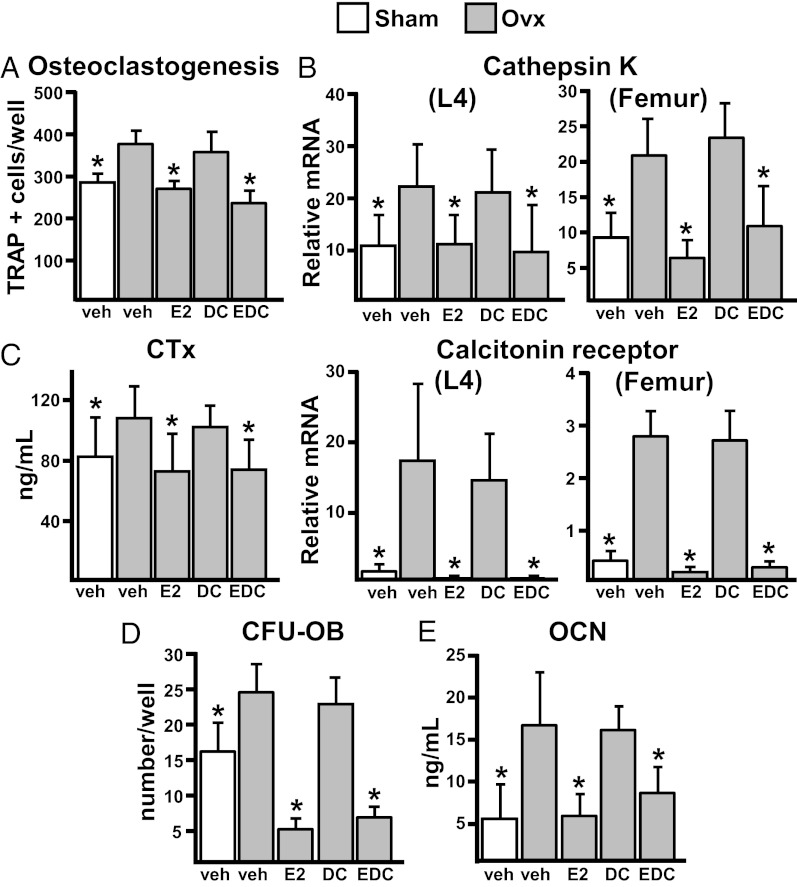
Figure 4.
Osteoclastogenesis and the increase in bone remodeling are prevented by EDC. A, Number of TRAP+ cells generated from bone marrow cells, pooled from the femurs of 3 mice per group and plated in triplicate cultures with M-CSF and RANKL for 5 days. B, Cathepsin K and calcitonin receptor mRNA levels by quantitative PCR in vertebral bone and femoral shafts (n = 8 animals per group). C, CTx levels in serum collected immediately before euthanasia (n = 10 animals per group). D, Bone marrow cells described in A were plated in triplicate at a density of 2 × 10⁶ cells/well. CFU-OBs were stained with von Kossa stain after 25 days to detect mineral. E, Osteocalcin (OCN) levels in serum collected immediately before euthanasia (n = 10 animals per group). Bars represent means and SD. *, P < .05 vs OVX, vehicle-treated by 1-way ANOVA. DC, empty dendrimer; veh, vehicle.

