Figure 1.
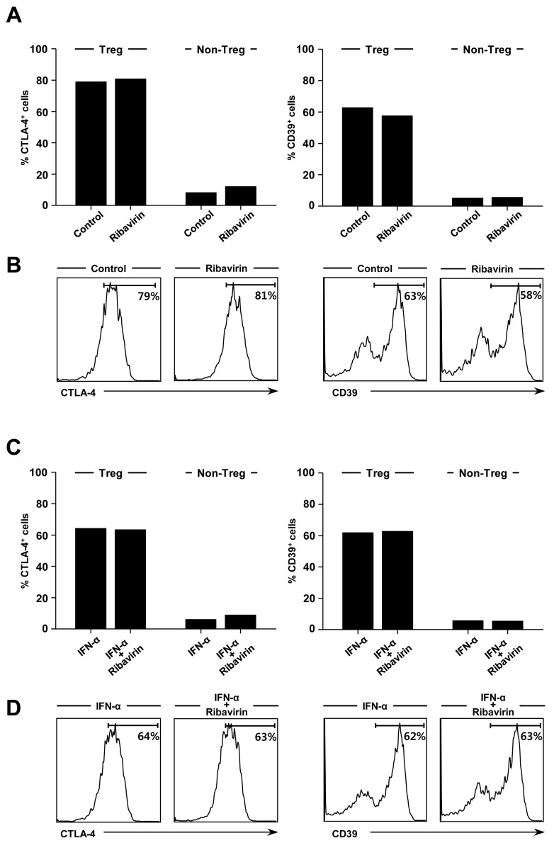
The effect of ribavirin on the expression of CTLA-4 and CD39. (A, B) PBMCs were isolated from whole blood of normal donors and treated with 2.5µg/ml of ribavirin for 3 days. The percentage of CTLA-4+ or CD39+ cells in Foxp3+CD4+CD25+ Treg cells and Foxp3-CD25- non-Treg CD4+ T cells was analyzed by flow cytometry (A). Representative histograms illustrate CTLA-4+ or CD39+ cells within the Foxp3+CD4+CD25+ Treg cell gate (B). (C, D) PBMCs were treated with 2.5µg/ml of ribavirin and 100 U/ml of IFN-α for 3 days. The percentage of CTLA-4+ or CD39+ cells in Foxp3+CD4+CD25+ Treg cells and Foxp3-CD25- non-Treg CD4+ T cells was analyzed by flow cytometry (C). Representative histograms illustrate CTLA-4+ or CD39+ cells within the Foxp3+CD4+CD25+ Treg cell gate (D). The experiment was performed with PBMCs of multiple donors, and data from a single donor is presented as representative data.

