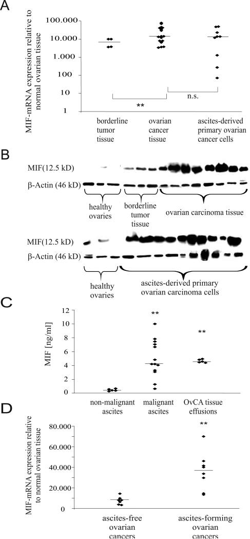
Figure 2.
MIF mRNA and protein are highly expressed in different OvCA entities. A, MIF mRNA expression was analyzed by quantitative real-time PCR in healthy ovaries (n=3), borderline tumor tissue (n=4), frozen tissue from OvCA (n=17) and in purified ascites-derived primary OvCA cells (n=9). MIF mRNA levels in tumors are expressed relative to the MIF expression found in normal ovaries. Note the logarithmic scale. Student's t-test was used to compare expression levels between tumors and healthy tissue and between tumors of different malignant potential (P < 0.05* and P < 0.01**). B, MIF protein levels were analyzed by Western Blot in 6 normal ovarian tissue samples, 10 lysates from ascites-derived primary OvCA cells, 7 tissue samples of solid OvCA and 3 tissue samples of borderline tumors. Equal loading was verified by b -actin staining. C, Soluble MIF was measured by ELISA in ascitic fluid from OvCA patients (n=13) and from patients with non-malignant ascites (n=5, p<0.01). Effusion of MIF from OvCA tissues was further assessed by placing 2 mg of freshly resected OvCA tissue in 2 ml of medium. Secreted MIF levels were determined by ELISA from the SN at 48 h. D, MIF mRNA levels were compared between ascites-free (n=7) and ascites-forming (n=8) OvCA.

