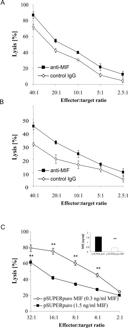
Figure 3.
Tumor-derived MIF suppresses lytic activity of NK cells. A, Primary human ascites-derived OvCA cells were labeled with 51Cr (NaCrO4), washed and used as targets (104/well) in a standard 4 h 51Cr-release assay with polyclonal NK cells as effector cells. Blocking anti-MIF antibody Mab289 or an isotype control antibody were used at 10 μg/ml. A representative of three assays performed in triplicates is shown (p<0.05 by ANOVA). B, SN were generated from 105 primary ascites-derived OvCA cells cultured for 72 h in 2 ml RPMI1640 medium with FCS and antibiotics. Polyclonal NK cells were pre-treated with these SN for 48 h in the presence of either blocking anti-MIF antibody Mab289 or an irrelevant isotype control (both used at 10 μg/ml). Subsequently, the NK cells were used in a 4 h 51Cr-release assay against the primary targets that had been used to generate the SN. A representative of 3 experiments is shown (p<0.01 by ANOVA) C, SK-OV-3 cells were transfected with a MIF shRNA plasmid (pSUPERpuroMIF) or the respective control vector (pSUPERpuro). Downregulation of MIF secretion was confirmed by ELISA (n=3). SN were added for 48 h to polyclonal human NK cells before these were used as effectors in a 4 h lysis assay against SK-OV-3 wild-type targets. Target cell lysis was determined by flow cytometric analysis of PKH-26 and CFSE-stained SK-OV-3 cells. A representative of 3 experiments is shown (p=0.01 by ANOVA).

