
Figure 3.
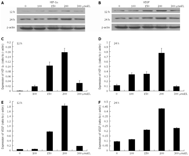
Protein expression levels of hypoxia-inducible factor-1α and vascular endothelial growth factor after exposure to 0-300 μmol/L CoCl2 for 12 and 24 h. A, B: The Western blotting analysis of protein expression levels of hypoxia-inducible factor-1α (HIF-1α) and vascular endothelial growth factor (VEGF); C, D: Histograms illustrating HIF-1α protein expression after exposure to various concentrations of CoCl2 for 12 h and 24 h; E, F: Histograms illustrating VEGF protein expression after exposure to various concentrations of CoCl2 for 12 h and 24 h.