Figure 2.

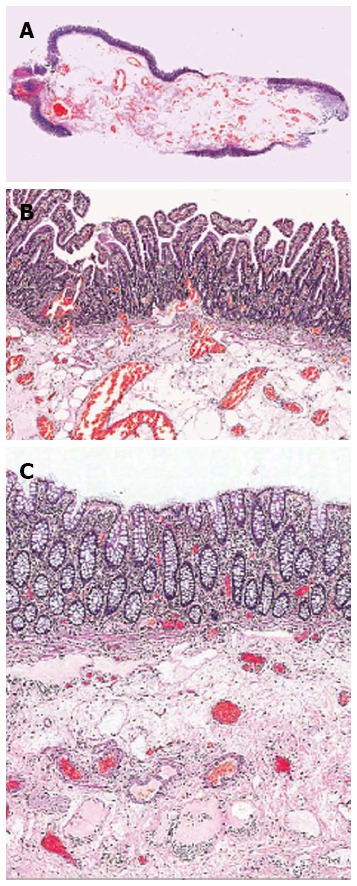
Histological section (hematoxylin and eosin staining). A: Case 1; B: Case 1 with normal small intestinal mucosal lining; C: Case 3 with normal large bowel mucosa overlying the submucosa which contains a prominent vascular component.

Histological section (hematoxylin and eosin staining). A: Case 1; B: Case 1 with normal small intestinal mucosal lining; C: Case 3 with normal large bowel mucosa overlying the submucosa which contains a prominent vascular component.