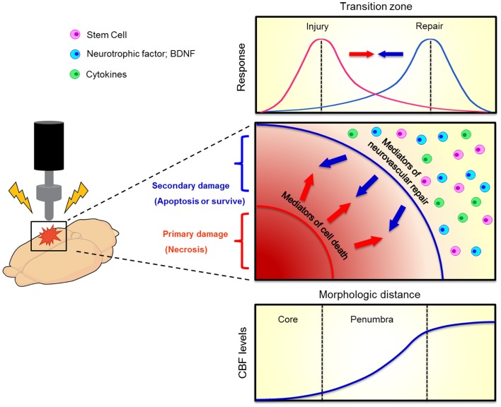
Figure 2.
Evolution of penumbra after TBI. The brain tissue surrounding the impacted core of TBI can become vulnerable to cell death due to spreading waves of pro-death cytokine mediators. This at-risk brain tissue corresponds to the penumbra which comprises the transition zone between injury and repair (top graphics). A therapeutic window exists for the repair process to abrogate the injury progression. When the brain cell faces damage, it suffers from two kinds of injuries, namely primary (necrosis) and secondary (apoptosis) cell death (middle graph). Neurovascular repair, such as transplantation of stem cells, upregulation of neurotrophic factors, and inhibition of pro-death cytokines, can rescue against the secondary cell death. The penumbra is traditionally defined as an area with mild to moderate reductions in cerebral blood flow (CBF, bottom graph). Such evolution of penumbra after brain injury was originally observed in stroke (Lo, 2008).

