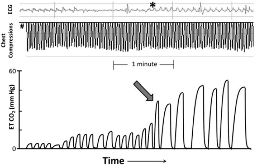
Fig. 2.
Using end-tidal (ET) CO2 to detect ROSC. From onset of arrest (#), note slow increase in end-tidal CO2 as compressions are delivered. With ROSC (arrow), organized ECG rhythm begins to appear under chest compression artifact (asterisk) and end-tidal CO2 rises suddenly to greater than 50 mmHg. Providers could have used the rapid rise in end-tidal CO2 as a clinical guide that there was a return of spontaneous circulation, without having to pause chest compressions and risk interruption of CPR for a rhythm check.

