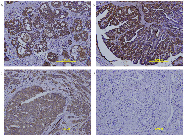
Figure 2.
Immunohistochemical staining for TFPI-2 expression in breast tumors. (A) A representative TFPI-2 positive staining image using sections of hyperplasia of mammary glands. (B) A representative TFPI-2 positive staining image using sections of intraductal papillomas. (C) A representative TFPI-2 positive staining image using sections of breast cancer. (D) A representative TFPI-2 negative staining image using sections of breast cancer. (magnification×200).

