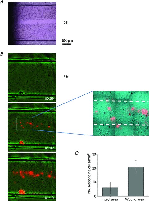
Figure 10. Enhanced ATP release responses during wound healing.

A, a ∼200 μm wide gap was created in a 2-day-old, fully confluent cell monolayer (t= 0) by removing a silicone strip that was attached to the bottom of the stretch chamber during cell seeding. The bright field image was taken with an inverted Nikon microscope, 20× objective. B, example of stretch-induced ATP release responses observed with cell cultures such as that appearing in A, after 16 h, when it was repopulated by new cells. The overlay of ATP-dependent luminescence (orange) and IR images (green) of cells in the stretch chamber are shown before stimulation, and 2 s and 10 s after 28% stretch. Note that almost all responses were within the wound-healing area, except for one artefact at the lower edge of the chamber, where some cells were agitated by debris visible in this spot on the image before stretch. The latter was excluded from the analysis. The zoomed region of the middle image discloses the ∼200 μm wide wound area, indicated by broken lines. Note that cells in the wound area are more spread out and flat compared to densely packed cells in the intact area, i.e. outside the broken lines. C, number of responding cells per mm2 in the wound-healing area was significantly enhanced when compared to the intact part of the same cell culture (P < 0.05, two-sample t test). Data are the average of five experiments/stretches with three different cell cultures.
