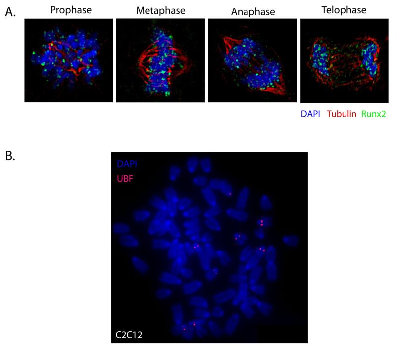
Figure 1. Metaphase Chromosome Spreads.
A. Runx2 is stable throughout mitosis. Synchronously growing Saos-2 cells were fixed and stained for DNA by using DAPI and for Runx2 by using a rabbit polyclonal antibody. Mitotic cells were identified by chromosome morphology. High-resolution images obtained by three-dimensional deconvolution algorithms reveal that Runx2 (green) is localized in mitotic chromosomes. A subset of Runx2 colocalizes with the microtubules, labeled by α-tubulin staining (red). B. A metaphase chromosome spread of mouse pre-myoblast C2C12 cells, immuno-labeled for Upstream Binding Factor (UBF; red) to identify nucleolar organizing regions (NORs).

