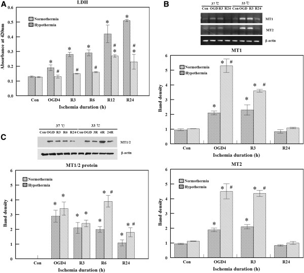
Figure 1.
Protective effect of hypothermia and induction of metallothionein (MT) gene expressions by hypothermia. (A) Lactate dehydrogenase (LDH) activity in the culture media was measured after bEnd.3 cells were subjected to 4 hours of oxygen glucose deprivation (OGD) and 24 hours of reperfusion (R) at 37°C or 33°C. LDH levels increased by OGD+R were diminished by hypothermia. Experiments were performed in triplicate. (B) Total RNA was isolated from bEnd.3 cells cultured in the normal control state, after 4 hours of OGD, and 3 to 24 hours after reperfusion initiation at 37°C or 33°C. The induction of MT expression peaked after OGD and decreased to basal levels during reperfusion. Hypothermia-augmented MT expression compared to normothermia. β-actin was used as the internal control. Band densities of the MT gene are expressed as the mean ± S.E.M. of five experiments. *P <0.05 compared to the control; #P <0.05 compared to normothermia. (C) MT protein expression was measured by western blot analysis. MT protein expression was induced after OGD and slowly declined during reperfusion. The level was higher with hypothermia. Band densities are expressed as the mean ± S.E.M. of five experiments. *P <0.05 compared to the control; #P <0.05 compared to normothermia.

