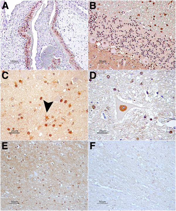
Figure 5.
CAPN6 immunostaining. (A) Staining of the sheep placental epithelium as a positive control for the technique. (B) Cerebellar cortex: moderate, diffuse neuropil staining of the gray matter of the molecular layer; neuronal cytoplasm was unstained. (C) Thalamus: in the white matter, the glial cells were strongly immunolabeled. Mainly, it consisted of nuclear staining, but the cytoplasm was occasionally observed (arrowhead). (D) Ventral horn of cervical spinal cord: Intranuclear staining was also evident in motor neurons. (E) Dorsal horn of a scrapie clinical case in which staining was stronger than in the (F) same region of a control sheep.

