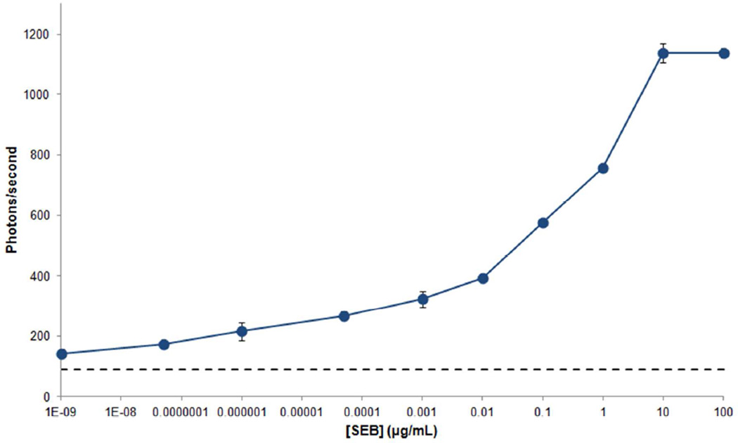
Figure 3.
Detection of SEB in buffer. Ten different concentrations of SEB dissolved in PBS were detected using a sandwich immunoassay: 10−9, 5×10⁻⁸, 10−6, 5×10⁻⁵, 10−3, 0.01, 0.1, 1, 10 and 100 µg/mL. LODs were determined from the mean plus three standard deviations of the background noise (dash lines). Error bars are based on the standard deviation of three replicates.

