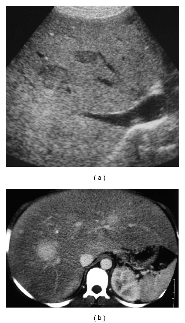
Figure 5.

Fourteen-year-old boy with glycogen storage disease type I and multiple HCA measuring from 1 to 3 cm diameter. (a) US shows an enlarged hyperechoic liver (steatotic) with well-delimited hypoechoic nodules. (b) CT performed at the arterial phase after contrast injection shows enlarged steatotic liver with multiple nodules that are strongly enhanced.
