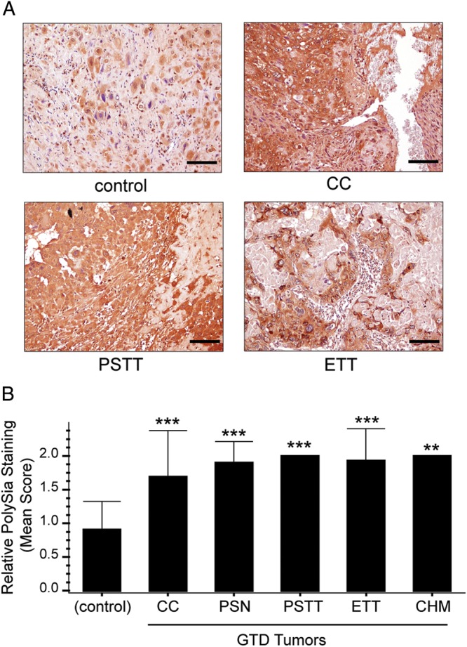
Fig. 6.

PolySia is overexpressed in biopsies of gestational trophoblastic disease (GTD) tumors. Biopsies of trophoblastic tumors and first-trimester normal placentas (controls) were paraffin-embedded and arranged into tissue microarrays. PolySia was detected with mAb 735 and visualized using DAB (brown). (A) Malignant tumors (choriocarcinomas [CC], placental site trophoblastic tumors [PSTT] and epithelioid trophoblastic tumors [ETT]) stained brightly for polySia compared with controls (normal first-trimester placental biopsies). Staining was not observed in necrotic regions. Scale bars, 40 µM. (B) The mean intensity of polySia immunoreactivity was significantly higher in biopsies from patients with GTD compared with controls, ***P < 0.001 or **P < 0.01. Error bars represent SD.
