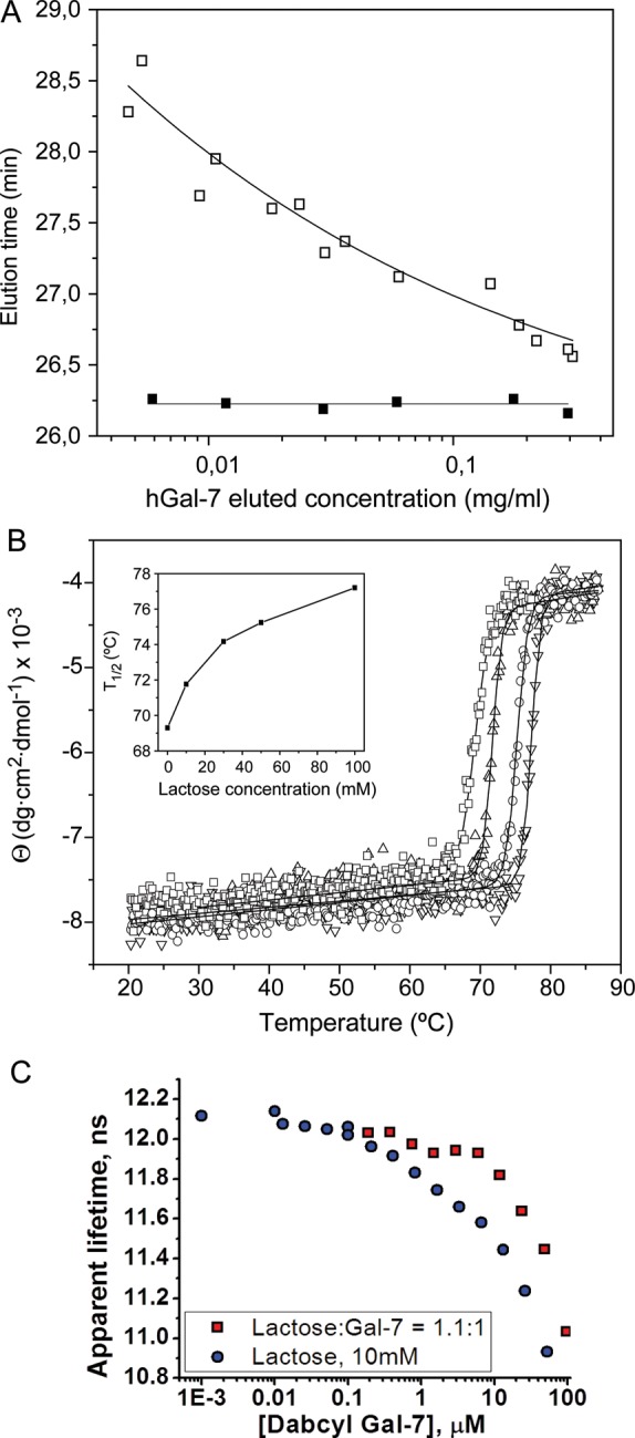
Fig. 7.

(A) Variation with protein concentration of the elution time of Gal-7 in gel filtration FPLC. 100 µL samples of Gal-7 at 0.04–2.4 mg/mL (2.6–160 μM monomer) were chromatographed in a Superose 12 column equilibrated at 30°C with 20 mM potassium phosphate buffer, pH 7.0, containing 2 mM DTT and 0.02% NaN3, in the absence (open squares) or presence of 10 mM lactose (filled squares). The concentration of the eluted protein was calculated using the dilution factor estimated for each peak from the ratio of width at half-height of the peak to the injection volume. (B) Effect of lactose on the stability of Gal-7 against thermal denaturation. The ellipticity at 220 nm of Gal-7 was monitored as a function of temperature, in the absence (open squares) and presence of 10 mM (open triangle), 50 mM (open circles) and 0.1 M (inverted triangle) lactose. The continuous lines correspond to the fit of a sigmoidal function to experimental data. Inset: Variation of the T1/2 with increasing lactose concentrations. (C) Hetero-FRET data showing the dependence of the apparent lifetime of donor fluorescence (fixed concentration of Edans-labeled Gal-7) as a function of the concentration of acceptor DABCYL-labeled Gal-7. The curve with red data points shows a molar ratio of lactose:Gal-7 of 1.1:1, and the blue data points are for lactose at 10 mM.
