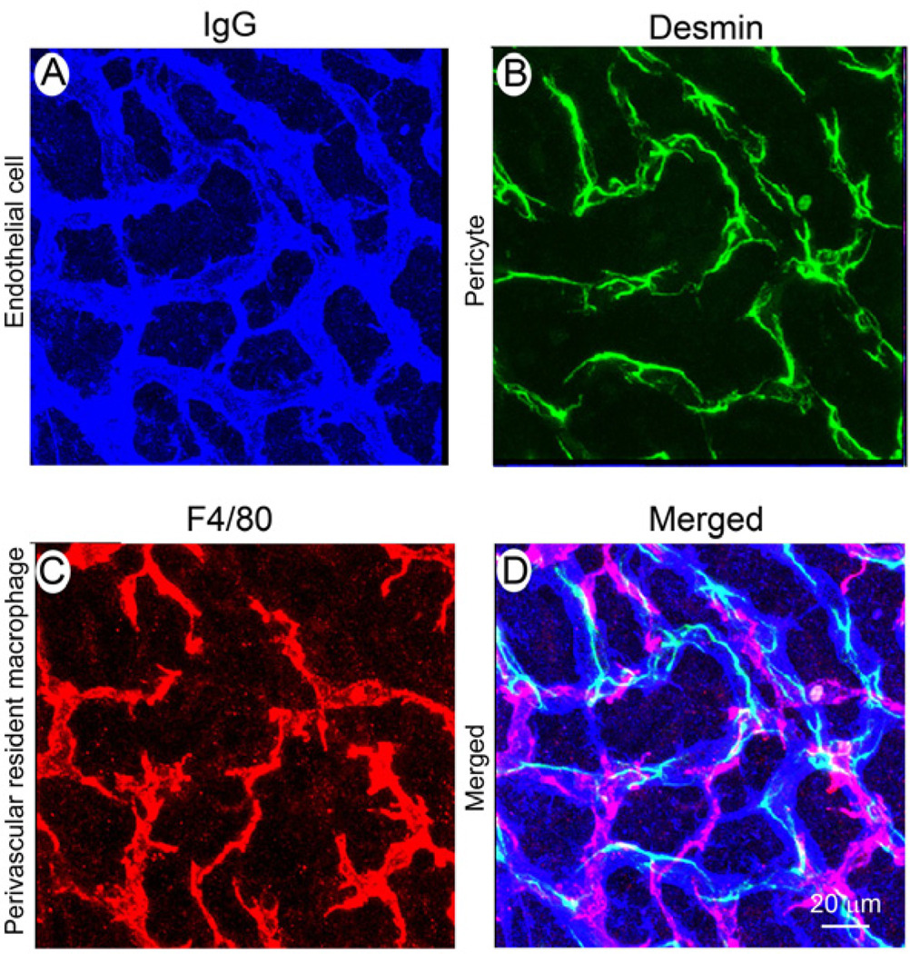
Figure 6. Cellular structure of the blood-labyrinth barrier.
Endothelial cells in normal BLB are identified with an antibody for mouse endothelial IgG (A, blue), pericytes with an antibody for desmin (B, green), and macrophages with an antibody for F4/80 (C, red). The merged image (D) shows the complexity of the blood-labyrinth-barrier.

