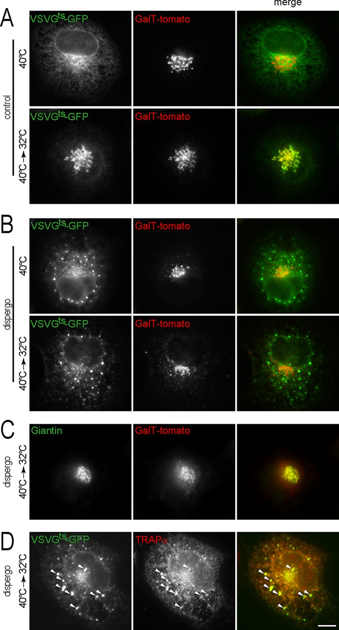
FIGURE 2:

Dispergo inhibits the traffic of VSVGts-GFP from the ER to the Golgi apparatus. BSC1 cells stably expressing GalT-tomato were transduced with adenovirus to express VSVGts-GFP overnight at 40°C. Cells were then treated with DMSO (control) or dispergo for 40 min at 40°C and subsequently shifted to 32°C for 20 min. (A) Control cells treated with DMSO only. (B–D) Cells treated with dispergo. Note in B the redistribution of VSVGts-GFP into patches in cells kept at 40°C during the dispergo treatment. (C) Under these experimental conditions, a significant amount of GalT-tomato colocalized with Giantin, validating the use of GalT-tomato as a Golgi marker. Endogenous Giantin was identified by immunostaining. (D) Colocalization of VSVGts-GFP patches with endogenous TRAPα, an ER marker (arrowheads) visualized by immunostaining in cells treated with dispergo. All images were from fixed BSC1 cells acquired using wide-field fluorescence microscopy. Scale bar, 10 μm.
