FIGURE 4:

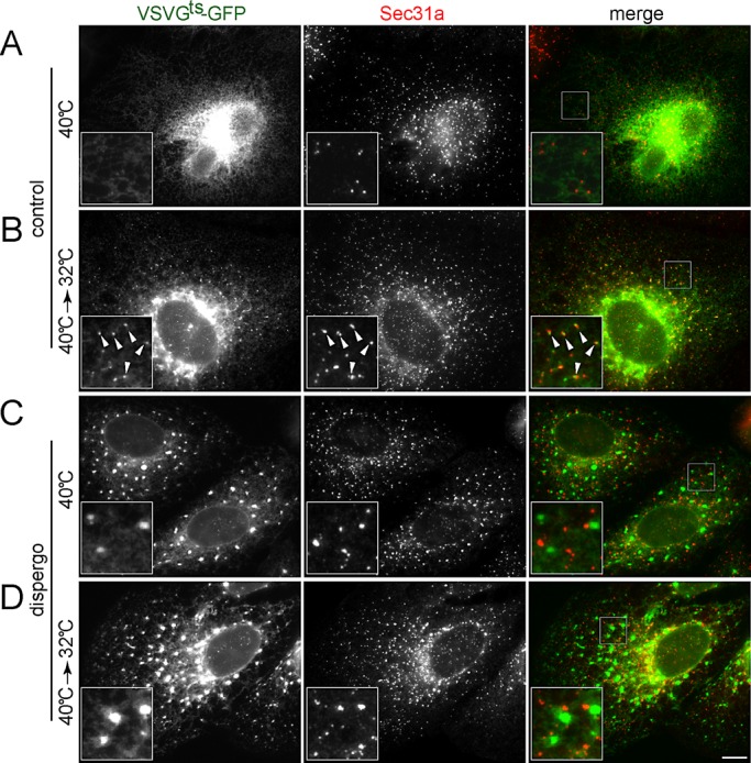
Dispergo blocks the recruitment of VSVGts-GFP to the ERES. BSC1 cells were transduced with adenovirus to express VSVGts-GFP overnight at 40°C. Cells were treated with either DMSO (control) or dispergo for 40 min (A, C). Subsequently, the temperature was shifted to 32°C for 10 min (40°C→32°C; B, D) before fixation and immunofluorescence detection of endogenous Sec31a. Images were acquired using wide-field fluorescence microscopy. The enlarged boxed regions in the merge images are shown in the bottom left of the corresponding composite images. Arrowheads highlight the colocalization of VSVGts-GFP with Sec31a in control. Scale bar, 10 μm.
