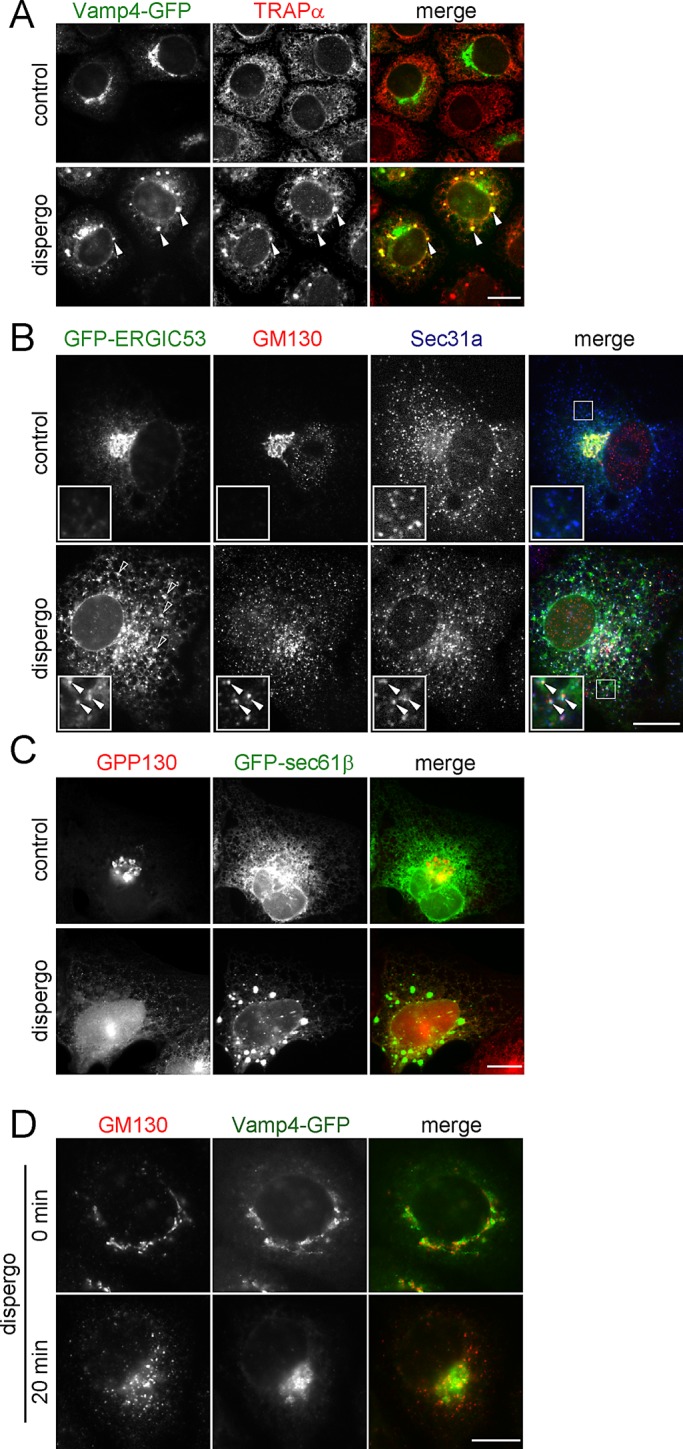
FIGURE 5:

Disappearance of the Golgi apparatus and retention of its components in the ER induced by dispergo. (A–C) Cells were treated for 3 h with DMSO (control) or dispergo before immunofluorescence labeling to follow the distributions of several ER (TRAPα, GFP-Sec61β, and Sec31a) and Golgi apparatus (VAMP4-GFP, GM130, ERGIC53-GFP, and GPP130) markers. (A) NRK cells stably expressing Vamp4-GFP immunostained with an anti-TRAPα antibody. Arrowheads highlight the colocalization of Vamp4-GFP and TRAPα in ER patches. (B) BSC1 cells transiently expressing GFP-ERGIC53 immunostained with antibodies specific for GM130 and Sec31a. The arrowheads in the enlarged boxed region highlight colocalization of GFP-ERGIC53 and GM130 at the ERES marked by Sec31a. Empty arrowheads indicate the ERGIC53-positive ER patches induced by dispergo. (C) BSC1 cells stably expressing GFP-Sec61β labeled with anti-GPP130 antibody. The images highlight lack of enrichment of GPP130 in ER patches. (D) Golgi markers had different kinetics of relocation upon dispergo treatment. NRK cells stably expressing Vamp4-GFP without or with dispergo treatment for 20 min were processed for immunofluorescence labeling of GM130. This example highlights that the redistribution of Golgi markers to the ER can occur at different rates (in this case GM130 was faster than Vamp4-GFP). All images were acquired by a wide-field microscope. Scale bars, 10 μm.
