FIGURE 6:
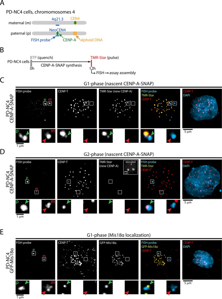
Timing of CENP-A assembly is maintained at neocentromeres. (A) Cartoon of maternal (canonical centromere) and paternal (neocentric) chromosome 4 in PD-NC4 cells. Indicated is chromosomal position 4q21.3, the site of neocentromere formation and the hybridization site of the FISH probe used. (B) Outline of quench-chase-pulse experiment in CENP-A–SNAP–expressing PD-NC4 cells. (C, D) Results of B for cells in G1 phase (C) or G2 phase (D), as indicated by nucleolar TMR staining, shown in rescaled inset. CENP-T indicates centromere positions. Enlargements display images of the hybridization sites of the FISH probe. Green arrows indicate the neocentromere, and red arrows show the homologous region on the maternal chromosome. (E) GFP-Mis18α–expressing PD-NC4 cells were stained for GFP and for 4q21.3 by FISH to detect Mis18α and the NeoCEN4, respectively. Enlargements as described above. Paternal (neocentric) and maternal 4q21.3 positions are indicated by p and m, respectively, in C–E.

