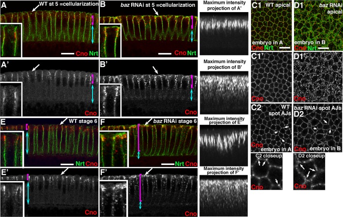
FIGURE 8:
Baz is not required for Cno assembly into spot AJs but does regulate precise Cno localization during polarity establishment. (A–D) Late cellularization. (A, B) Apical-basal cross-sections. (C1, D1) Apical surface sections of embryo in A and B. (C2, D2) Surface sections at level of normal spot AJs. In WT (A, C), Cno localizes along the apical end of the lateral membrane (A′, bracket) to spot AJs (C2). In maximum-intensity projections of multiple apical–basal sections (A′), cables of Cno that localize to tricellular junctions are apparent. Cno is also largely removed from the apical surface during cellularization (A′, E′, arrows). Reducing Baz by RNAi (B, F) leads to Cno spreading more basally (B′, bracket, vs. A′, bracket), to loss of organized Cno cables (B′, maximum-intensity projections), and to failure to exclude Cno puncta from the apical membrane (B′, arrow, C1′ vs. D1′), but Cno still continues to assemble into spot AJs (D2). (E, F) Gastrulation onset (stage 6). In WT, Cno remains in spot AJs (E′, bracket), and the apical–basal cables at tricellular junctions become even more prominent (E′, projections). baz RNAi leads to spread of Cno basally (F′), perturbs assembly of Cno cables at tricellular junctions (F′ maximum-intensity projection), and allows Cno puncta to accumulate at the apical surface. (F′, arrow and inset). Scale bars, 10 μm.

