FIGURE 8:
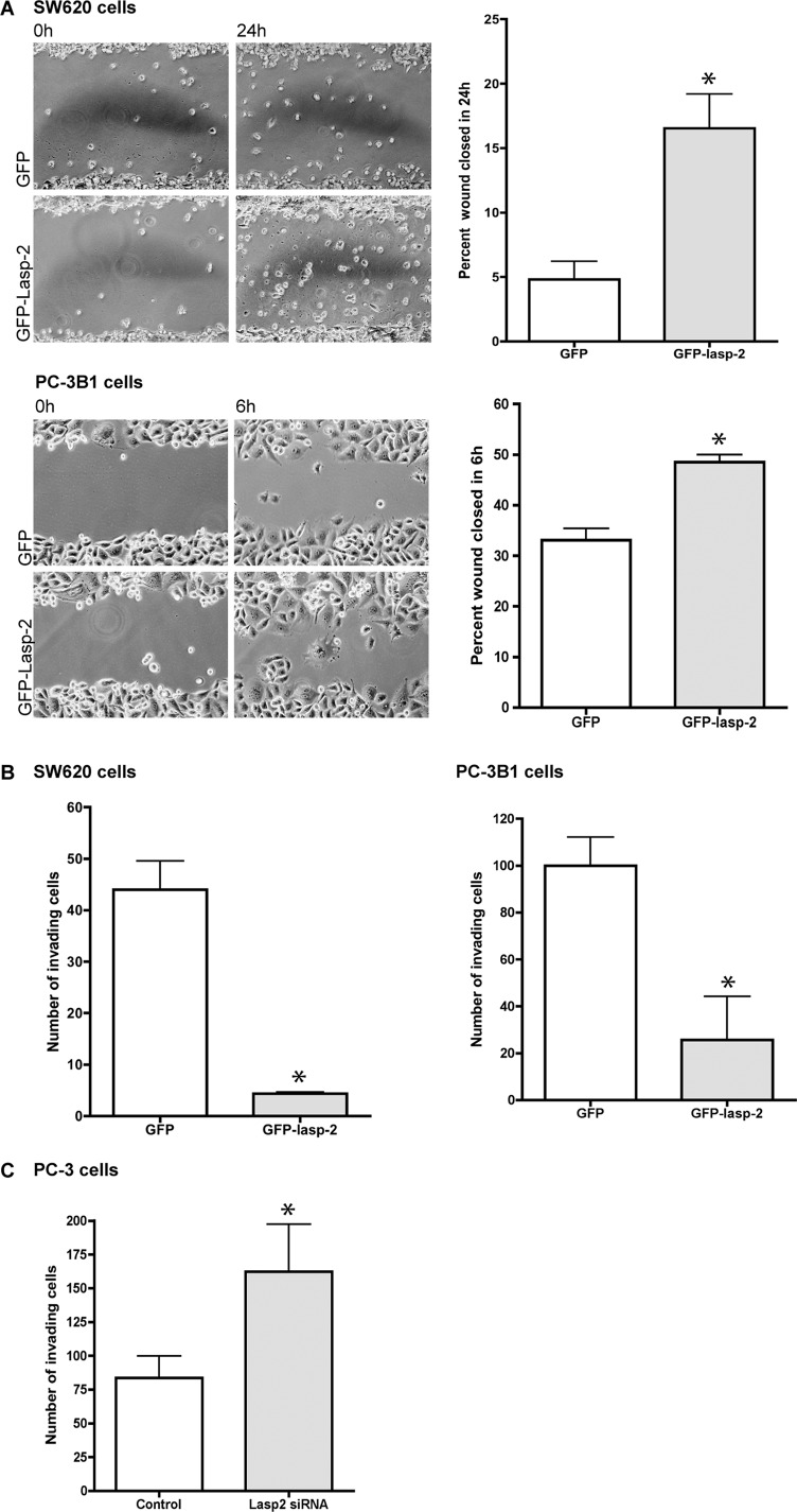
The overexpression of lasp-2 in SW620 and PC-3B1 cells increases cell migration rates but reduces cell invasion. (A) SW620 cells, derived from metastatic colorectal cancer, and PC-3B1 cells, derived from metastatic prostate cancer, migrated at a faster rate when GFP–lasp-2 is expressed. There was a threefold increase in the wound-healing rate in cells expressing GFP–lasp-2 in the SW620 cells. There was a 1.4-fold increase in the wound-healing rate in cells expressing GFP–lasp-2 in the PC-3B1 cells. *p < 0.05. (B) Cell invasion is reduced in cells expressing GFP–lasp-2. GFP–lasp-2–expressing cells invaded the chamber an average of 11-fold less than control cells in SW620 cells and invaded the chamber an average of fourfold less than control cells in PC-3B1 cells. *p < 0.005. (C) Loss of lasp-2 protein leads to an increase in cell invasion. Two different siRNA sequences to human lasp-2 were used to reduce lasp-2 protein levels in PC-3 cells. Cells with lasp-2 protein knocked down invaded the chamber approximately twofold more than controls. Data from one of the siRNA sequences are shown. *p < 0.05.

