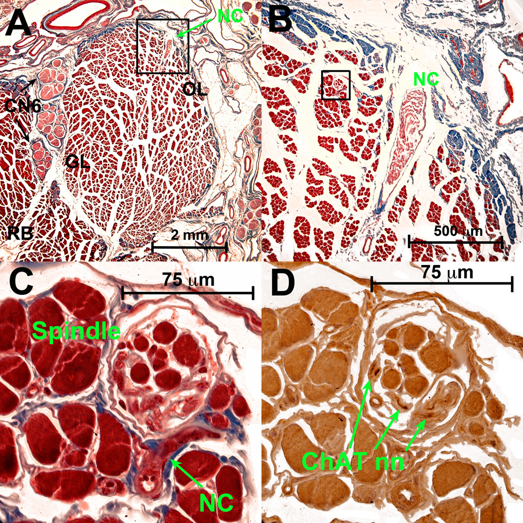
Fig. 3.
Bovine LR muscle. A. Low power Masson trichrome stain shows classical CN6 that has repeatedly divided on the global (left) LR surface as it courses anteriorly to enter on the global surface (not shown) of the global layer (GL). Nonclassical (NC) n. enters LR orbital layer (OL) from its superior orbital surface in the deep orbit. B. Magnified view of region in black square shown in A, demonstrating entry of NC n. C. High power view of region in black square shown in B, demonstrating NC n. entry into a spindle, distinct from the blood vessel seen below it. D. ChAT in nearby serial section demonstrates dark brown immunoreactivity of some axons in NC nerve in the spindle shown in C. RB – retractor bulbi muscle. Other abbreviations as in Fig. 1.

