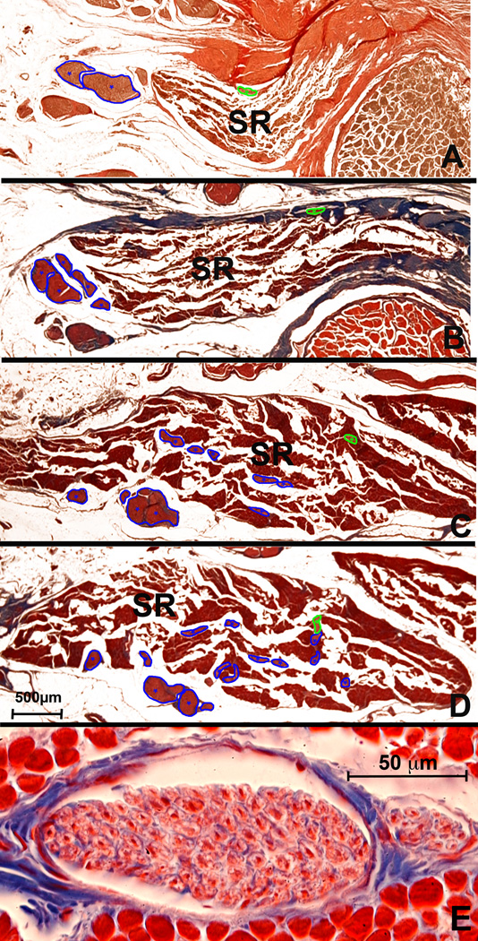
Fig. 4.
Human superior rectus (SR). Panels A – D from posterior to anterior at low power. Classical motor n. from superior division of CN3 (marked in blue), enters the EOM but is joined by a ChAT positive NC n. (marked in green) entering the SR more posteriorly through its orbital surface. E. High power cross section of anterior portion of NC nerve and its smaller branch (right) showing predominance of large fibers. A. Van Gieson stain. B – E. Masson trichrome stain. 500 µm scale bar for A – D. 50 µm scale bar for E.

