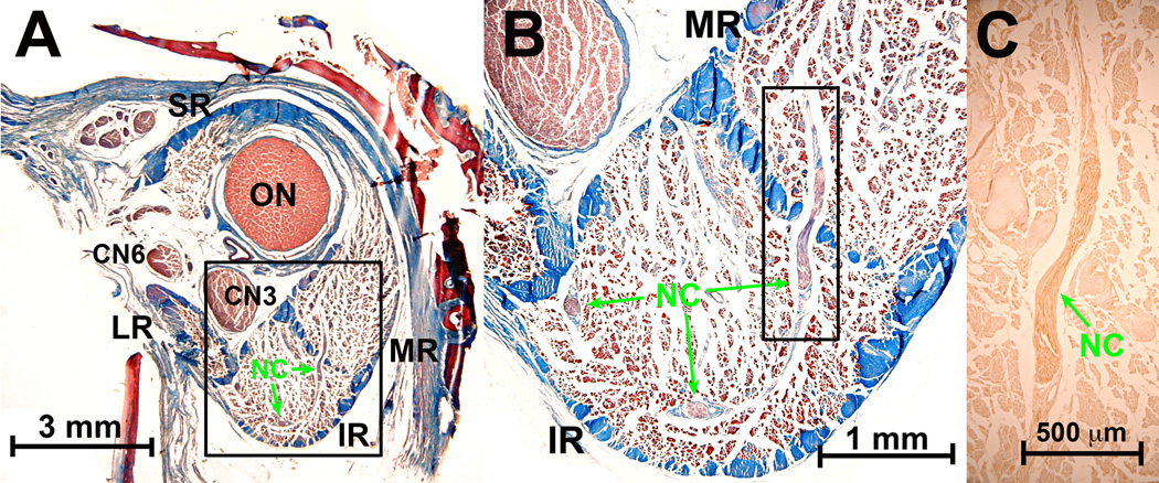
Fig. 5.
Apex of human orbit near the rectus origin at the annulus of Zinn, 37.81 mm posterior to corneal surface. A. Masson trichrome shows n. branch that appeared in deep sections and traveled from inferior rectus (IR) to medial rectus (MR) muscle. A small branch from this n. entered the main trunk of CN3 before CN3 gave any branch to any EOM. B. Higher magnification of region in A outlined by rectangle demonstrates that NC n travels through the collagenous septum separating the IR ad MR. C. Darker brown central ChAT immunostaining in nearby serial section of region demarcated by rectangle in B demonstrates positive brown staining of NC n. Abbreviations as in Fig. 1.

