
Fig. 4. cyp26b1 mutants and morphants display deficiencies in midline cartilages of the neurocranium and visceral skeleton.
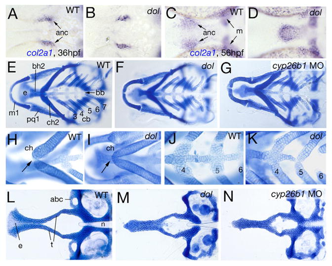
All panels show ventral views of zebrafish head regions. (A–D) col2a1 in situ hybridization at indicated stages. (E–N) Alcian Blue stainings of cartilaginous craniofacial elements at 120 hpf. Pharyngeal arches are numbered (1, mandibulare; 2, hyoid; 3–7, branchial/gill arches 1–5). (E–G) Overviews of visceral skeleton. (H–K) Magnified views of ceratohyals (H, I) or pharyngeal arches 4–6 (J, K). Arrows in H, I point to ceratohyal (ch) attachment in midline. (L–N) Flat-mounts of neurocranium, revealing the absence of medial ethmoid (e) and anterior basicranial commissure (abc) in mutant and morphant. anc, chondrocytes of anterior neurocranium; bb, basibranchial; bh, basihyal; cb, ceratobranchials; m, Meckel’s cartilage; n, notochord; pq, palatoquadrate; t, trabeculae cranii.