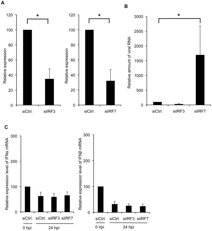
Figure 5. Enhancement of viral propagation with siIRF7 is not mediated by inhibition of type I IFN induction.
(A) The total RNA from siRNA-transfected MDCK cells was isolated and subjected to quantitative real-time RT-PCR. (B) The siRNA transfected MDCK cells were infected with PR8 virus at a MOI of 0.03. The amount of viral RNA in culture supernatant was determined by quantitative real-time RT-PCR. Data are shown as fold change in amounts of viral RNA from siRNA-transfected cells compared with that from control cells. (C) The expression of mRNA for each IFN-α/β was measured at indicated time points after infection by quantitative real-time RT-PCR. Data are shown as fold change in expression of mRNA for each IFN-α/β compared with that of control siRNA-transfected cells and normalized to the values for 18S rRNA. The data are representative results of three independent experiments. Asterisks indicate statistically significant differences compared with the control (*P<0.05).

